
What Causes Hyperdontia? (Genetics, Syndromes, and Treatments)

Dr. Emrah YEŞİLYURT
Dr. Emrah Yeşilyurt is the Founder of Avangart Clinic. He combines advanced dental expertise with a genuine commitment to helping patients feel comfortable and informed about their oral health journey.
Finding an extra tooth can be concerning, whether it appears as a tooth behind a tooth, an extra tooth in the gums, or a tooth growing high in the upper jaw. This condition is called hyperdontia, which means having supernumerary teeth in addition to the normal number of baby or adult teeth.
Hyperdontia is not usually dangerous in itself, but it should never be ignored. Extra teeth can be hidden under the gums or grow into the mouth, interfering with the dental arch, tooth eruption, bite alignment, or appearance.
The exact cause of hyperdontia is not always clear, but it is most strongly linked to genetic and developmental factors. In many cases, hyperdontia appears to run in families or occurs alongside certain syndromes, while in others it may be related to overactivity in the dental lamina during tooth development.
This is why two patients with an extra tooth may have very different experiences. For example, a child may have a hidden mesiodens blocking a front tooth from erupting properly, while an adult may not realise there is an extra tooth until an X-ray is taken for crowding, discomfort, or cosmetic concerns.
- Medical Disclaimer: The information here is provided for general educational purposes and should not be used as a substitute for professional dental advice, diagnosis, or treatment. Hyperdontia can vary greatly from one patient to another, and proper assessment requires a clinical examination supported by appropriate imaging. If you are concerned about an extra tooth or an unusual eruption pattern, we recommend booking a consultation for an accurate diagnosis and personalized treatment plan.

The Primary Causes of Hyperdontia (Supernumerary Teeth)
When patients ask us what the main hyperdontia causes are, the honest answer is that there is not always one single explanation. In most cases, we look at two likely factors: genetics and changes that happen during early tooth development.
The first is heredity. Some people seem more likely to develop extra teeth because of the way tooth formation is passed through the family. That does not always mean there is a serious underlying condition, but it does tell us that genetics can play an important role.
The second is developmental. During early formation, teeth begin from a tissue called the dental lamina. If that tissue becomes unusually active, it may create an extra tooth bud instead of stopping at the usual number.
One patient may have a single hidden tooth with no obvious family history, while another may have several extra teeth and a clearer inherited pattern.
In the clinic, this difference matters. If there is a clear family history or signs of a wider syndrome, we look beyond the extra tooth itself and assess the case more thoroughly than we would with a simple isolated finding.
Genetic Syndromes Associated with Hyperdontia
In some cases, what seems like an extra tooth in the gums is not just an isolated dental finding. It can be part of a wider genetic condition that affects how the bones, jaws, and teeth develop. When that pattern is present, we look at the full clinical picture rather than treating the tooth as a one-off anomaly.
Cleidocranial Dysplasia
Cleidocranial dysplasia is one of the clearest genetic conditions linked to hyperdontia. It affects bone development, especially in the skull and collarbones, and it often has a strong effect on how teeth form and erupt.
In these patients, teeth may stay trapped in the jaw for longer than expected, come through late, or appear in unusual numbers and positions. It is not uncommon to see a mix of retained baby teeth, unerupted adult teeth, and crowding all at once.
Gardner Syndrome
Gardner syndrome is another genetic condition that can be associated with hyperdontia. It is linked to a higher rate of dental anomalies, including extra teeth, impacted teeth, and other irregularities in the jaws.
This syndrome is also associated with cysts and abnormal tissue growths elsewhere in the body. That is why unusual dental findings may sometimes be an early clue to a wider medical condition, rather than an isolated issue.
Cleft Lip and Palate
Cleft lip and palate can disrupt normal development in the upper jaw and dental arch. When the tissues in this area do not join in the usual way during early development, the teeth near the cleft may erupt in unusual positions, fail to erupt, or develop in excess.
This is one reason some patients present with what feels like a tooth growing on the roof of the mouth or close to the palate. In these cases, the position of the tooth often reflects the way the surrounding bone and soft tissues formed.
Fabry Disease & Ehlers-Danlos Syndrome
Fabry disease and Ehlers-Danlos syndrome are less common associations, but they are still relevant. Fabry disease can have an impact on several body systems and oral structures. Ehlers-Danlos syndrome affects connective tissue and can affect dental development in more complex ways.
These conditions do not account for most cases of hyperdontia, but they remind us that extra teeth can sometimes be part of a broader inherited pattern. If a patient already has one of these diagnoses, dental assessment should always be planned with that wider medical context in mind.
Types of Supernumerary Teeth and Where They Grow
| Type | Typical location | What it may look like clinically | Primary clinical risks |
|---|---|---|---|
| Mesiodens | Between or just behind the upper front teeth | May appear as an extra front tooth, a tooth behind another tooth, or may stay hidden under the gum | Delayed eruption of a front tooth, crowding, displacement of nearby teeth, midline spacing issues |
| Paramolar | Beside a molar, usually towards the cheek or tongue side | May sit slightly outside the normal dental arch and can be easy to miss if small or partially erupted | Plaque retention, cleaning difficulty, local gum irritation, crowding |
| Distomolar | Behind the last molar, often near the wisdom tooth region | Usually found at the very back of the mouth and may remain unerupted | Impaction, pressure on nearby teeth, difficulty cleaning the area, cyst-related changes if impacted |
A mesiodens near the front of the mouth raises different concerns from a distomolar at the back, even though both are forms of hyperdontia.
Location is only part of the picture, though. Some supernumerary teeth look quite similar to normal teeth, while others are smaller, more pointed, or irregular in shape. That combination of position and shape helps us understand how the tooth is likely to behave and whether it is likely to interfere with normal development.
Signs and Complications: Why Extra Teeth Are a Problem
One of the confusing things about hyperdontia is that it does not always announce itself clearly. Many patients have no pain at all, and the extra tooth is only discovered when an X-ray is taken for another reason. That is why hyperdontia symptoms can range from obvious crowding to no noticeable symptoms whatsoever.
When problems do happen, it is usually because the mouth simply does not have room for an extra tooth. Teeth are expected to come in a certain order, in very particular positions. So one extra tooth can throw that pattern off.
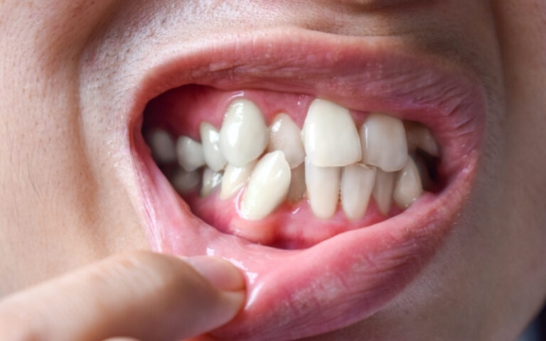
Some of the most common complications we see are:
Crowding
An extra tooth can press against the surrounding teeth and leave everything looking cramped. Over time, this can make the smile look uneven and create tight areas that are much harder to keep clean.
Impaction
Sometimes the extra tooth blocks another tooth from coming through properly. In that situation, the normal tooth may remain trapped beneath the gum and fail to erupt into its proper position.
Displacement of nearby teeth
A nearby tooth may still erupt, but not where it should. It might come through at an angle, twist slightly, or shift out of line because the extra tooth has taken up the space it needed.
Cyst formation
In some cases, a sac of fluid can form around an unerupted tooth. This is one reason the answer to whether hyperdontia is dangerous is sometimes more complex than a simple yes or no. The condition itself is often manageable, but the complications should not be ignored.
You may also notice delayed eruption, gaps that do not make sense, or one tooth appearing to come through while another seems missing. In children, this is often the first clue that something beneath the gums is affecting normal development.
How is Hyperdontia Diagnosed?
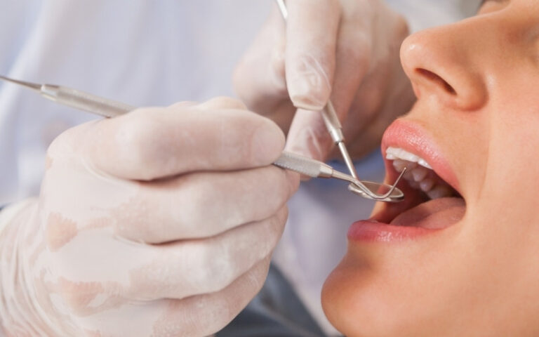
Diagnosing hyperdontia starts with a careful clinical examination, but the full picture usually comes from imaging. Some extra teeth are visible during a routine check-up, while others are completely hidden under the gums and only appear on a scan.
In most cases, we diagnose hyperdontia in three steps:
Visual examination
We first look at the teeth that are visible, the spacing between them, and whether the eruption pattern looks normal for the patient’s age.
Panoramic X-ray
This gives us a broad view of the upper and lower jaws in one image. It is often the most useful first scan for finding impacted supernumerary teeth, checking their position, and seeing how close they are to nearby roots.
CBCT scan when needed
If the tooth is in a difficult position or close to important structures, Cone Beam Computed Tomography gives us a far more detailed 3D view. This helps us plan safely and accurately, especially before any surgical or orthodontic decision is made.
Routine dental examinations are important because hyperdontia often goes unnoticed in its early stages. A child may come in because a front tooth is late to appear, or extra teeth growing in adults may only be discovered during an assessment for crowding or alignment.
What matters most is not just confirming that an extra tooth is there, but understanding exactly where it is, how it is sitting in the jaw, and whether it is affecting other teeth. That is what allows us to decide whether simple monitoring is enough or whether the case needs more active management.
Treatment Options for Hyperdontia
The best hyperdontia treatment depends on what the extra tooth is actually doing in your mouth. Not every supernumerary tooth needs to be removed. If it’s not impacting on neighbouring teeth, crowding, or interfering with normal development, then it may be most sensible to monitor it over time.
If the tooth is not causing pressure, crowding, delayed eruption, or hygiene problems, careful observation may be all that is necessary. In these cases, we review the area periodically and use follow-up imaging to make sure the tooth is not starting to create a new problem.
When treatment is needed, the most common next step is removal. This is usually planned as a surgical extraction, especially if the tooth is impacted, positioned deep in the jaw, or close to neighbouring roots. The exact approach depends on the tooth’s location, the patient’s age, and how much influence it is having on the rest of the mouth.
In many cases, removing the extra tooth is only the first stage. If the surrounding teeth have shifted, rotated, or failed to erupt properly, orthodontic treatment may then be needed to bring everything back into a healthier position. This may involve braces or clear aligners, depending on the case and the amount of movement required.
For example, a child may have an adult front tooth that cannot come through properly because an extra tooth is blocking it. After the extra tooth is removed, the adult tooth may erupt by itself, although some children still need orthodontic help to bring it into place.
That is why treatment planning is individual rather than automatic. We look at whether the tooth can be monitored, whether oral surgery is the better option, and whether follow-up orthodontic care will be needed to restore proper alignment and bite function.
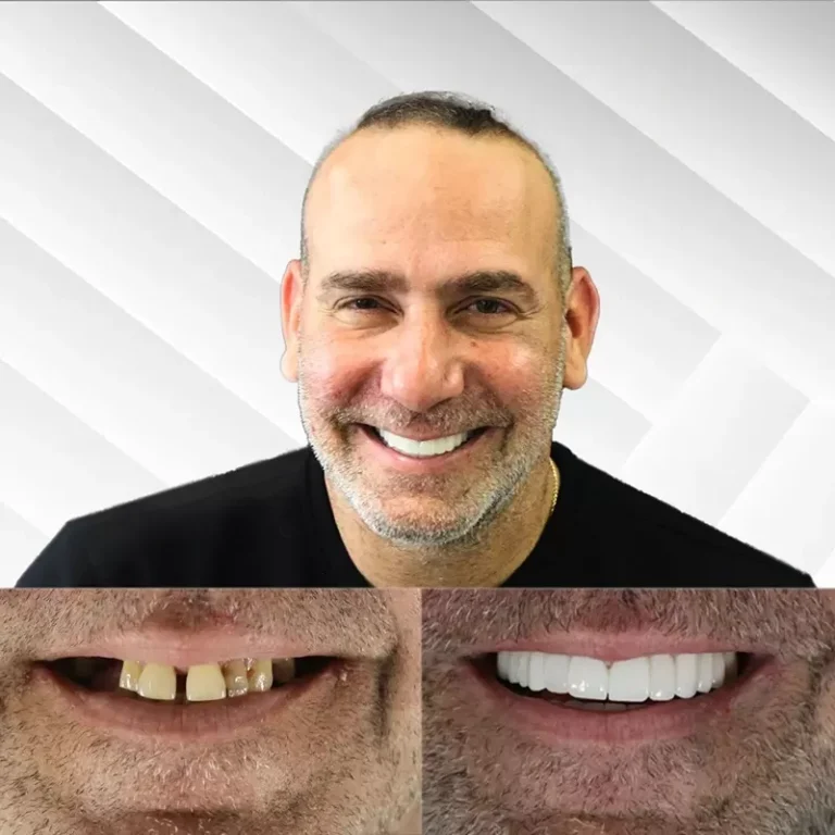
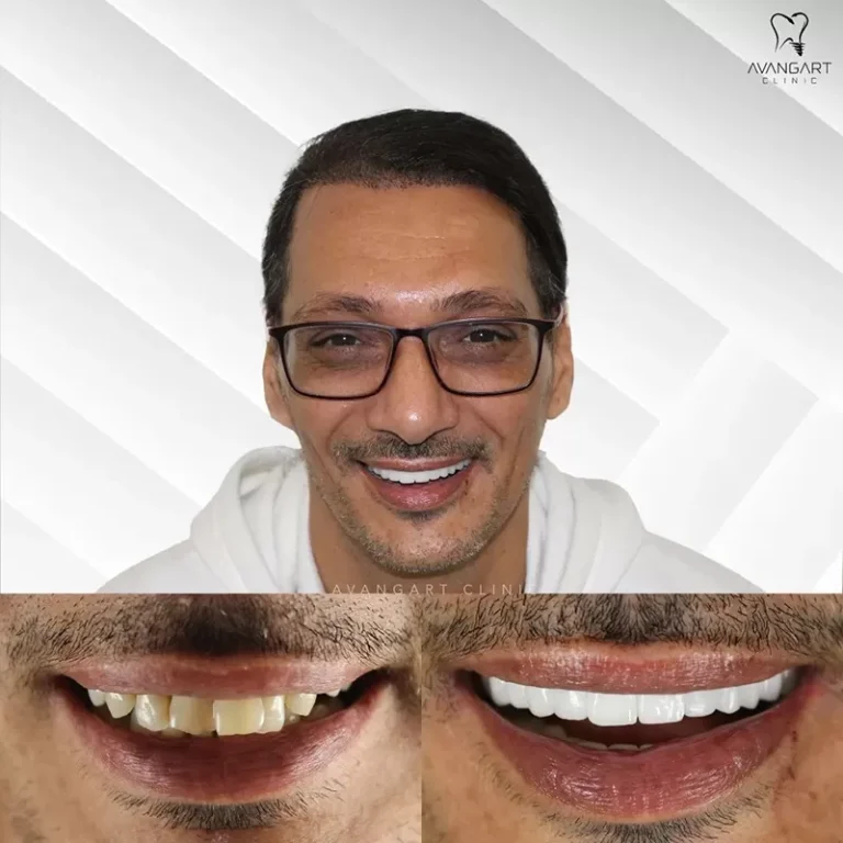
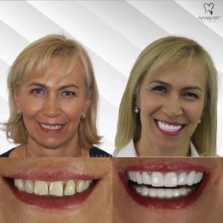
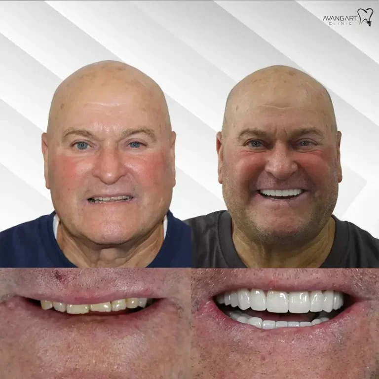
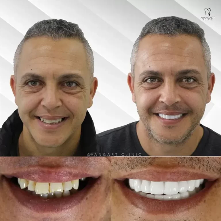
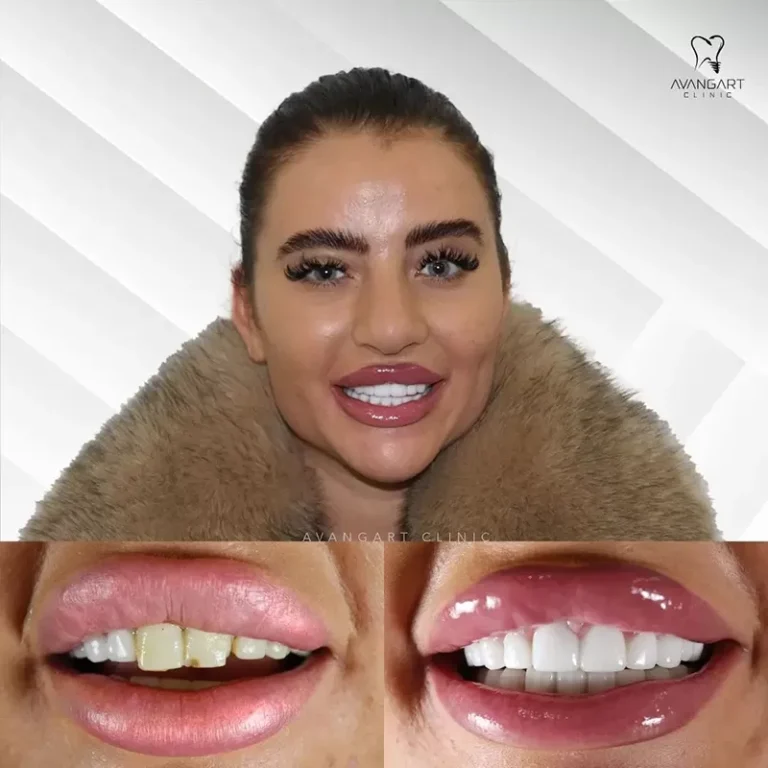
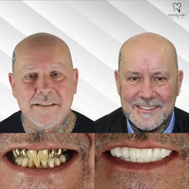
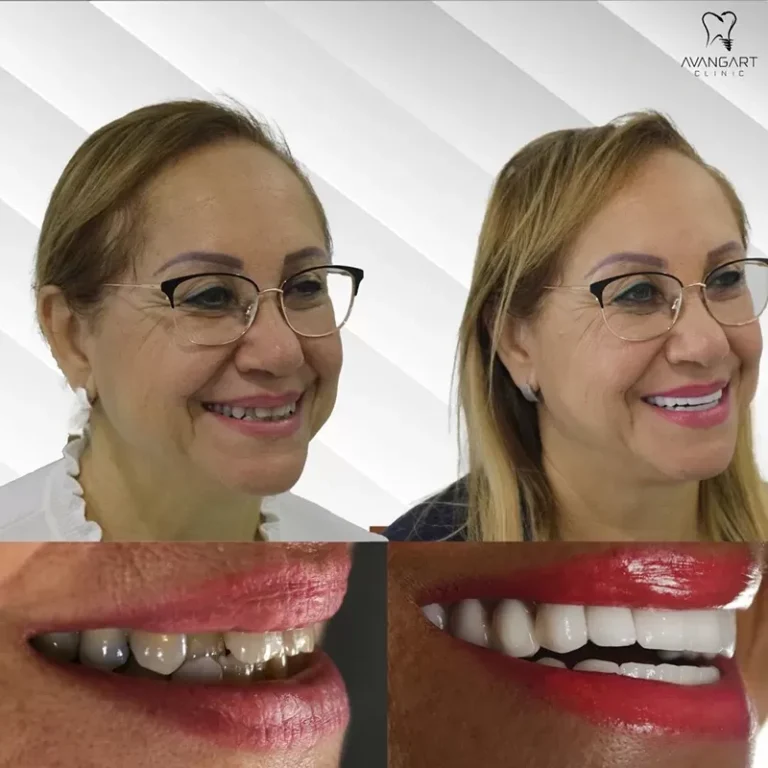
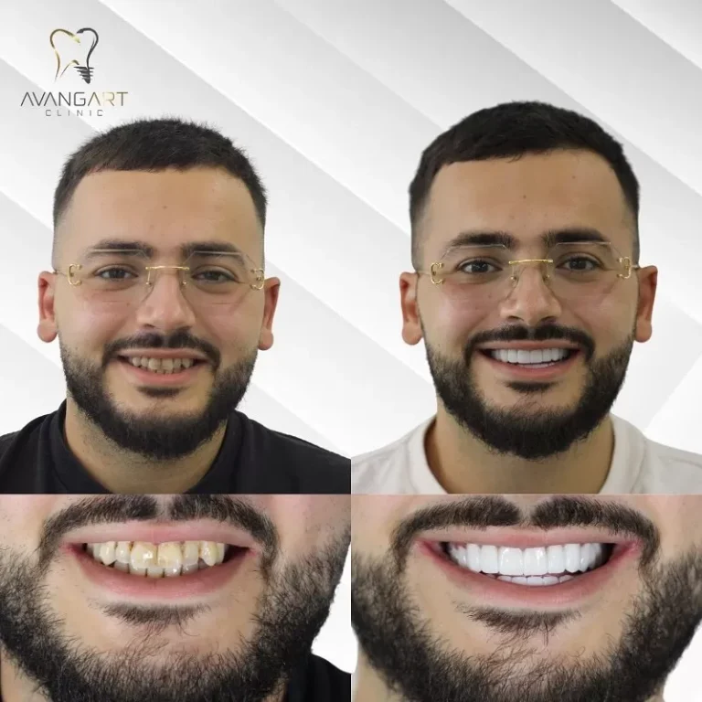
Before and After: Transformative Results with Avangart Clinic
We carry out all our dental treatments with care and strive to give your smile an elegant appearance.
FAQs
Yes — hyperdontia is relatively uncommon. It affects about 3.8% of adult teeth and 0.6% of baby teeth. We do see it in practice, but it is still unusual enough to need proper dental investigation.
It can, although many people feel nothing at all. Pain usually happens when the extra tooth presses on nearby teeth, irritates the gums, or causes inflammation.
No. Some supernumerary teeth can simply be monitored if they are not disturbing anything around them. At Avangart, we usually recommend removal when the tooth is blocking eruption, pushing other teeth out of place, or creating a higher risk of cysts, crowding, or hygiene problems.
Most of the time, an extra tooth develops because the tissues that make teeth were overactive during early development. Genetics can also play a part, and that’s why some cases appear to be familial. It is not caused by anything you did wrong.
It is not considered normal to have an extra tooth growing from the gum, but it is a recognised dental condition. Sometimes, patients first notice a small tooth-like shape appearing in an unusual place. When that happens, an examination and X-ray are the best next steps.
Hyperdontia can look very different from one person to another. It may appear as a small extra tooth between the front teeth, a tooth erupting behind another, or a tooth appearing in an unusual part of the upper jaw. In other cases, it is completely hidden and only seen on imaging.
Adults can be diagnosed with hyperdontia, but the extra tooth usually forms earlier and is only discovered later on an X-ray or during treatment planning.
That depends on where the extra tooth is and whether it is affecting anything around it. Some remain quiet and stable, but others can push nearby teeth out of place, interfere with the bite, make cleaning harder, or contribute to cyst formation. Even without pain, it is worth having it assessed properly.
Worried About Extra Teeth? Schedule a Consultation
If you have noticed an unusual tooth in your own mouth, or your child seems to have a tooth coming through in the wrong place, it is worth having it checked sooner rather than later. In many cases, the next step is simply a diagnostic X-ray and a careful consultation to understand exactly what is happening beneath the surface.
With the right assessment, we can explain what is happening and talk you through the most appropriate next steps.
Our specialized clinical team, led by Oral & Maxillofacial Surgeon Dr. Emrah Yeşilyurt and Prosthodontist Dr. Egemen Aras, works with international patients to plan treatment clearly and safely.
If you are traveling from the US, the UK, or Europe, you can securely email your digital panoramic X-rays or CBCT scans ahead of time for a preliminary remote evaluation. To speak with our international patient care coordinators, contact us at +90 212 211 82 11 or email contact@avangartclinic.com.